
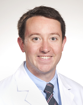
Arno H. Fried, MD, FACS, FAANS
Pediatric & Adult Neurosurgeon
An arteriovenous malformation (AVM) is a rare condition that often has no symptoms for many years. Some people don’t know they have an AVM until they experience a seizure, a severe headache, or another neurological problem. In other cases, it is found during a brain scan performed for an unrelated reason.
An unexpected AVM diagnosis can be overwhelming, but understanding your condition is the first step toward making informed treatment decisions.
At Advanced Neurosurgery Associates, we provide expert evaluation and treatment for adults with AVMs at our four New Jersey locations: Rutherford, Jersey City, New Brunswick, and Morristown. Our team works with you to explain your diagnosis, discuss your treatment options, and develop a personalized care plan based on your needs.

An arteriovenous malformation (AVM) is an abnormal connection between arteries and veins. Normally, blood flows through tiny blood vessels called capillaries before reaching the veins. In an AVM, this step is skipped.
As a result, blood flows through the vessels at higher pressure. This can weaken the blood vessels over time and increase the risk of bleeding in the brain or spinal cord.
Most AVMs are present at birth and develop before a baby is born. However, many people do not have symptoms during childhood.
Symptoms often appear between the ages of 35 and 40. In some cases, an AVM is only discovered after it causes a health problem or during imaging for another condition.
A brain AVM can increase the risk of a hemorrhagic (bleeding) stroke, especially in younger and middle-aged adults.
Early evaluation by an experienced neurosurgeons of new jersey can help:
Visual suggestion:
Include a diagram comparing normal blood flow with blood flow through an AVM to help patients understand how the condition affects the brain.
Meet Our Expert Team
At ANA — Movement. Vitality. Medicine., we are committed to providing exceptional pain, vein, and wellness services that empower individuals to reclaim their health and well-being. With our team of Harvard-trained experts, we strive to deliver transformative outcomes that restore comfort, confidence, and quality of life. ANA stands for our mission to help every patient relieve discomfort, restore motion, and renew life.

Arteriovenous malformations (AVMs) can affect both men and women. They occur in people of all ethnic backgrounds.
Most AVMs never cause symptoms. However, when symptoms do appear, they can lead to serious neurological problems if left untreated.
Women should know that pregnancy increases blood volume and blood pressure. These changes may trigger symptoms in a previously undiagnosed AVM. If you have an AVM or develop new neurological symptoms during pregnancy, seek medical evaluation promptly.
Arteriovenous malformation symptoms in adults vary considerably depending on whether and where bleeding has occurred. Many adults are entirely asymptomatic. When symptoms do arise, they may include:
Certain diagnostic tests may be performed to determine if surgery would be an effective treatment. This surgical evaluation can involve a number of tests to develop the diagnosis of the cause and location of the epileptic seizures.
Evaluation prior to surgery for epilepsy has changed radically in the past few decades, most notably since the advent of long-term video-electroencephalography (EEG) monitoring in the late 1970s, and advanced neuroimaging, specifically with the additional use of MRI.
To develop the epilepsy diagnosis before surgery, patients partake in extensive testing, such as:
It is also important to consider the emotional effects. Neuropsychology and psychiatry are a vital part of ANA’s epilepsy team’s evaluation.
Electroencephalography (EEG) is the fundamental measure of identifying seizure focus. The epileptologists on our team are adept at reading the spikes and sharp waves that constitute a spectrum of shifting seizure focus. Most patients undergoing evaluation for surgery will require EEG with video telemetry to document the seizure pattern.
However, an appropriate focus goes beyond single readings. Our experts evaluate EEG monitoring over a period of time. This is a critical measure to determine a clear onset and location of seizure activity.
While computerized tomography (CT) scan can provide some useful information about lesions which may be causing seizures, the “gold standard” for locating and evaluating brain lesions is magnetic resonance imaging (MRI). The greatest advance of MRI technology for epilepsy is the incredible clarity of both the normal and pathological anatomy of the brain. Since the mid-1980s, this technology has allowed us to routinely identify lesions that were previously undetectable.
Nuclear medicine, which deals with imaging the body for both diagnostic and treatment purposes, plays an important role in the pre-surgical assessment of patients with refractory epilepsy. This is evident in the case of single-photon emission computed tomography (SPECT) which is used to determine the seizure onset zone.
SPECT (single proton emission computed tomography) has an important role in the investigation of surgical candidates. Ictal (meaning taking place during a seizure) SPECT has also been useful to study seizure-spread patterns. Blood flow increases in the brain area in which the seizure originates and the blood flow can become less than normal in that same area during non-seizure states. By injecting a small and safe amount of a radioactive substance into a patient’s blood stream via IV we can evaluate the blood flow in the brain at a given time. Changes in blood flow during and after a seizure can be helpful in localizing seizure focus and spread patterns.
Diagnosing an arteriovenous malformation (AVM) starts with a detailed medical history and neurological examination. Your neurosurgeon will review your symptoms and recommend imaging tests to confirm the diagnosis.
Your evaluation may include:
These imaging studies help your neurosurgeon:
Visual suggestion: Add an infographic showing the diagnostic process: Neurological Exam → MRI → Cerebral Angiography (DSA) → CTA/MRA (if needed) → Personalized Treatment Plan.
For many people, medication is the first step in controlling epilepsy. Taking your medication exactly as prescribed gives you the best chance of reducing or preventing seizures.
Some patients become seizure-free with their first medication. Others may need a different medicine or a combination of medications to achieve better seizure control.
Finding the right medication can take time. Your doctor will closely monitor your response to treatment and make changes if needed.
During follow-up care, you may have:
Our goal is to find the safest and most effective treatment plan to help you manage epilepsy and maintain your quality of life.
A ketogenic diet is a special meal plan that is high in fat and low in carbohydrates. Instead of using sugar for energy, the body burns fat. This change can help reduce seizures in some children with epilepsy.
The ketogenic diet is often recommended when medications do not fully control seizures. It is carefully planned and monitored by a healthcare team to make sure your child gets the right nutrition.
Many children see fewer seizures after starting the diet.
Possible benefits include:
The results are different for every child.
The ketogenic diet should only be started under medical supervision. Your child’s care team will monitor growth, nutrition, and overall health with regular follow-up visits.
Our epilepsy specialists will determine whether the ketogenic diet is the right treatment option and support your family throughout the process.
Treatment decisions for adult AVMs are among the most nuanced in all of neurosurgery. Not every AVM requires immediate intervention, and the risks of treatment must always be weighed carefully against the risks of observation. Our team uses the Spetzler Martin Grading System — the standard clinical tool for assessing AVM surgical risk — to evaluate each patient’s case based on AVM size, location, and venous drainage pattern before any treatment recommendation is made.
Open surgical removal through a craniotomy remains the most definitive treatment, offering immediate cure when complete resection is achieved. Most appropriate for accessible, lower-grade AVMs — particularly those that have already bled. Our surgeons use intraoperative neurophysiological monitoring and image guided navigation throughout the procedure.
Focused radiation — delivered via systems such as Gamma Knife — gradually obliterates the AVM over two to three years without open surgery. Best suited for smaller, deep seated AVMs that carry high surgical risk. Radiosurgery does not provide immediate protection against hemorrhage
during the obliteration period, a consideration that factors into treatment timing discussions.
A catheter-based procedure used to selectively block blood vessels feeding the AVM, reducing its size and blood flow. Most commonly used as a preparatory step before surgery or radiosurgery rather than a standalone cure, though in select cases it can achieve significant AVM reduction.
For older adults, patients with small asymptomatic AVMs, or those with high-grade lesions where treatment risks outweigh benefits, careful monitoring with serial imaging may be the most appropriate path. Observation is an active, managed decision — not inaction.
Epilepsy surgery is measured by the level of improvement in seizures and quality of life. Adult studies of epilepsy surgery have shown that seizures can be greatly reduced or totally controlled in some cases, and many can stop AEDs. Pediatric studies also report that the majority of infants and young children show favorable outcomes in seizure control, and can also stop AEDs after surgery.

If you have been diagnosed with an arteriovenous malformation (AVM) or have symptoms that may be related to an AVM, getting the right care is important. An experienced neurosurgeon can review your imaging, explain your diagnosis, and discuss the best treatment options.
At Advanced Neurosurgery Associates, we provide specialized AVM care at our four New Jersey locations in Rutherford, Jersey City, New Brunswick, and Morristown.
We believe every patient deserves personalized care. Our team takes the time to answer your questions, explain your treatment options in clear language, and create a care plan that fits your needs and goals.
An AVM diagnosis doesn’t have to be overwhelming. Contact Advanced Neurosurgery. Associates today for expert evaluation and a clear path forward.
Common arteriovenous malformation symptoms may include headaches, seizures, dizziness, muscle weakness, numbness, vision problems, and difficulty speaking. Symptoms vary depending on the AVM’s location and size.
You should seek arteriovenous malformation treatment if you experience severe headaches, seizures, bleeding, or neurological symptoms. Early diagnosis and treatment can help reduce the risk of complications.
Arteriovenous malformation treatment options may include minimally invasive embolization, microsurgery, radiosurgery, or a combination of treatments depending on the condition and severity.
Advanced Neurosurgery Associates provides specialized arteriovenous malformation treatment in New Jersey with advanced diagnostic imaging and personalized care plans.
Arteriovenous malformation specialists are neurosurgeons and vascular experts trained to diagnose and treat AVMs using advanced surgical and minimally invasive techniques.
Look for experienced arteriovenous malformation specialists in Rutherford, NJ who offer comprehensive neurological care, modern treatment options, and personalized patient support.
Advanced Neurosurgery Associates offers expert arteriovenous malformation treatment in Rutherford, New Jersey, using advanced technology, individualized treatment plans, and compassionate care.
Yes, minimally invasive arteriovenous malformation treatment in Rutherford, New Jersey, may include embolization and radiosurgery to help reduce recovery time and improve outcomes.
Some arteriovenous malformations can be completely treated or removed depending on their size and location. Your specialist will recommend the best treatment approach after evaluation.
You can contact Advanced Neurosurgery Associates to schedule a consultation with experienced arteriovenous malformation specialists in Rutherford, NJ, for diagnosis and treatment planning.