If you or your child has been diagnosed with Chiari malformation, understanding the specific type is an important step toward choosing the right treatment and knowing what to expect. Although the condition shares a common name, there are four recognized Chiari malformation types, each with different anatomical features, symptoms, and levels of severity.
Some people experience mild symptoms that can be managed with observation, while others require specialized neurosurgical care to relieve pressure on the brain and restore normal cerebrospinal fluid (CSF) flow. The type of Chiari malformation often determines when symptoms appear, how the condition progresses, and whether surgery may be recommended.
This guide explains the different Chiari malformation types, highlights the differences between Type 1, Type 2, Type 3, and Type 4, and provides an overview of diagnosis, treatment, and when to seek care. Whether you’re researching for yourself or your child, understanding these distinctions can help you make informed healthcare decisions.
At Advanced Neurosurgery Associates, our experienced pediatric and adult neurosurgeons provide comprehensive evaluation and personalized treatment for patients with Chiari malformation using advanced diagnostic imaging and evidence-based care.
What Are the Types of Chiari Malformation?
Chiari malformation is generally described in four types: Type 1, Type 2, Type 3, and Type 4. Type 1 is the most common and may not be diagnosed until later childhood or adulthood. Type 2 is typically present at birth and is commonly associated with myelomeningocele, a severe form of spina bifida. Type 3 is rare and more severe. Type 4 refers to an underdeveloped cerebellum and is also very rare.
The type of Chiari malformation, a patient’s symptoms, and MRI findings help doctors determine whether monitoring or treatment may be appropriate.
What Is Chiari Malformation?
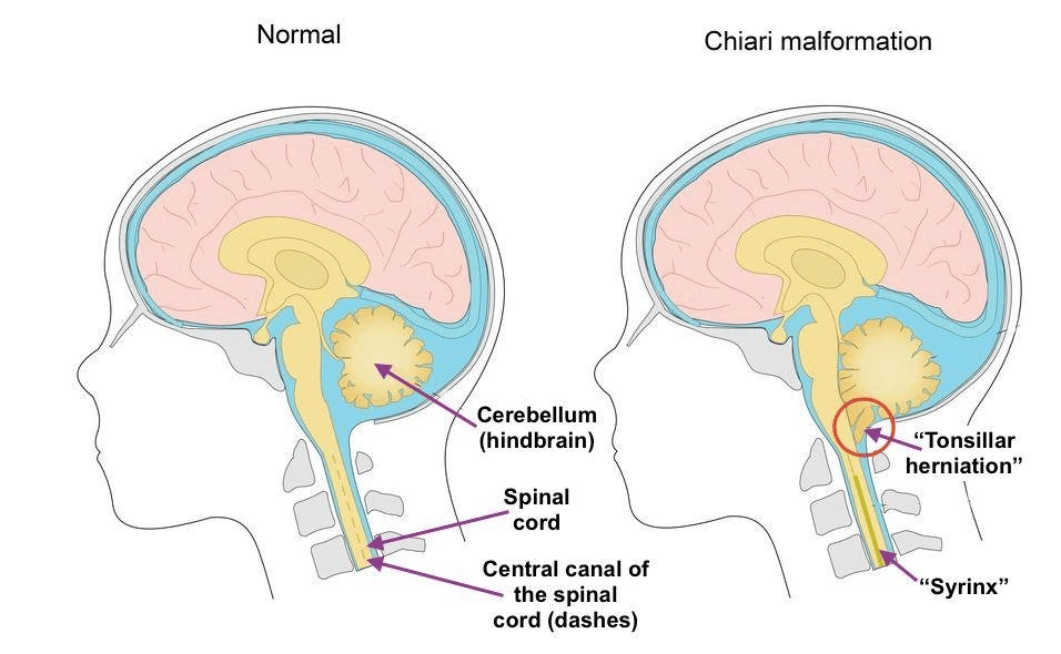
Chiari malformation is a structural abnormality in which part of the cerebellum extends through the opening at the base of the skull into the spinal canal. This displacement can interfere with the normal flow of cerebrospinal fluid (CSF), leading to pressure on the brainstem and upper spinal cord.
Most Chiari malformations are congenital, meaning they develop before birth. However, symptoms may not appear until later in childhood or adulthood, particularly in patients with Chiari malformation Type 1.
Common symptoms include headaches, neck pain, dizziness, poor balance, numbness, muscle weakness, swallowing difficulties, and vision changes. The severity of symptoms depends on the type of Chiari malformation and the degree of pressure on surrounding neurological structures.
Comparison of Chiari Malformation Types
| Type | Usually Diagnosed | Severity | Common Association |
| Type 1 | Older children & adults | Mild–Moderate | Syringomyelia |
| Type 2 | Before birth or infancy | Moderate–Severe | Spina bifida |
| Type 3 | At birth | Severe | Encephalocele |
| Type 4 | At birth | Very Severe | Cerebellar hypoplasia |
Chiari Malformation Type 1
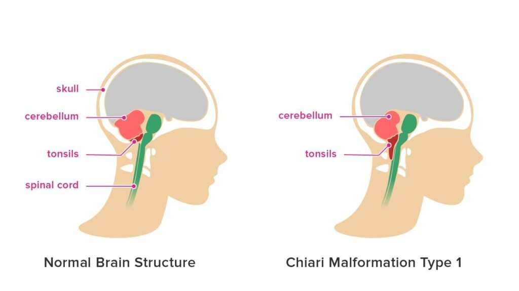
Chiari malformation Type 1 is the most common form and is often diagnosed during adolescence or adulthood. In this type, the cerebellar tonsils extend into the upper spinal canal, but the brainstem usually remains in its normal position.
Some people have no symptoms, while others develop headaches, neck pain, dizziness, balance problems, numbness, muscle weakness, or difficulty swallowing. Symptoms often become more noticeable over time or may worsen with coughing or straining.
Many patients with mild symptoms can be monitored with regular neurological examinations and MRI scans. When symptoms become severe or cerebrospinal fluid flow is affected, surgery may be recommended to relieve pressure and improve neurological function.
Chiari Malformation Type 2
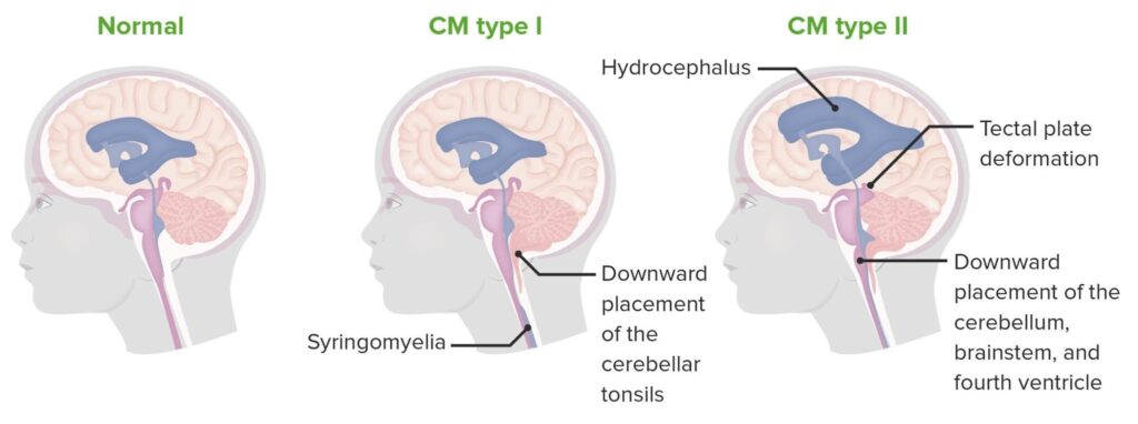
Chiari malformation Type 2 is a congenital condition that is almost always associated with myelomeningocele, the most severe form of spina bifida. In this type, both the cerebellum and part of the brainstem extend downward through the opening at the base of the skull.
Children with Type 2 may experience feeding difficulties, breathing problems, weakness, developmental delays, or hydrocephalus. Because this form is more complex than Type 1, treatment often involves a multidisciplinary team that may include pediatric neurosurgeons, neurologists, and rehabilitation specialists.
Early diagnosis and ongoing follow-up play an important role in improving long-term outcomes.
Chiari Malformation Type 3
Chiari malformation Type 3 is an extremely rare and severe form of the condition. In this type, part of the cerebellum and sometimes the brainstem protrude through an opening in the back of the skull, a condition known as an encephalocele. Because of these significant structural abnormalities, Type 3 is usually diagnosed before birth through prenatal imaging or immediately after delivery.
Infants with Chiari malformation Type 3 often experiences serious neurological complications, including developmental delays, muscle weakness, coordination difficulties, and breathing or feeding problems. The severity of symptoms varies depending on the amount of brain tissue involved.
Treatment typically involves specialized neurosurgical care to repair the skull defect and manage associated neurological conditions. Although Type 3 is uncommon, early diagnosis and coordinated care can help improve overall quality of life and long-term management.
Chiari Malformation Type 4
Chiari malformation Type 4 is the rarest and differs from the other forms because the cerebellum is underdeveloped (cerebellar hypoplasia) rather than displaced into the spinal canal. This condition develops during fetal growth and is associated with significant neurological impairment.
Children with Type 4 may experience severe developmental delays, movement disorders, poor muscle tone, and coordination difficulties. Since the condition affects brain development itself, treatment focuses on supportive care, rehabilitation therapies, and ongoing neurological management rather than correcting the structural abnormality.
Chiari Malformation: Type 1 vs Type 2
Although Chiari malformation Type 1 and Type 2 share similarities, they differ in how they develop, when symptoms appear, and the conditions they are associated with.
| Feature | Type 1 | Type 2 |
| Age at Diagnosis | Older children, teens, or adults | Before birth or infancy |
| Brain Structures Involved | Cerebellar tonsils | Cerebellum and brainstem |
| Associated Conditions | Syringomyelia, scoliosis | Spina bifida, hydrocephalus |
| Severity | Mild to moderate | Moderate to severe |
| Treatment | Observation or surgery | Often requires multidisciplinary care and surgery |
Type 1 is the most common form and may remain symptom-free for years. In contrast, Type 2 is a congenital condition that is frequently diagnosed early in life because of its association with spina bifida and other neurological abnormalities.
How Symptoms Differ in Adults and Children
The symptoms of Chiari malformation often depend on both the type of malformation and the patient’s age.
Adults
Adult Chiari malformations are most commonly diagnosed with Chiari malformation type 1 and may experience the following:
- Persistent headaches, especially after coughing or straining
- Neck pain
- Dizziness or vertigo
- Balance and coordination problems
- Numbness or tingling in the arms or legs
- Muscle weakness
- Difficulty swallowing
- Vision changes or tinnitus
Children
Children may present with symptoms such as the following:
- Feeding difficulties
- Developmental delays
- Poor coordination
- Breathing or swallowing problems
- Frequent headaches
- Weakness in the arms or legs
- Sleep apnea
- Scoliosis
Because symptoms can overlap with other neurological conditions, a thorough evaluation by an experienced neurosurgeon is essential.
Related Reading: Learn more about the signs and symptoms on our Chiari Malformation Symptoms page.
How Is Chiari Malformation Diagnosed?
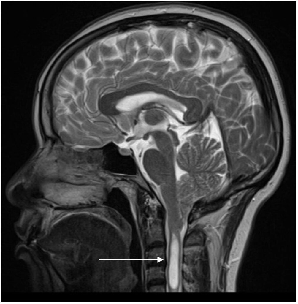
Diagnosing Chiari malformation begins with a detailed medical history and neurological examination. If Chiari malformation is suspected, your neurosurgeon may recommend imaging studies to confirm the diagnosis and determine the specific type.
Common diagnostic tests include:
- MRI (Magnetic Resonance Imaging): The most accurate test for evaluating the brain, spinal cord, and cerebrospinal fluid flow.
- Cine MRI: Used to assess how cerebrospinal fluid moves around the brain and spinal cord.
- Neurological Examination: Evaluates balance, reflexes, muscle strength, sensation, and coordination.
These evaluations help determine whether monitoring or surgical treatment is the most appropriate option.
Treatment Options

Treatment depends on the type of Chiari malformation, symptom severity, and MRI findings. Some patients require only regular monitoring, while others benefit from surgical treatment.
Common treatment options include:
- Observation with routine neurological follow-up
- MRI monitoring
- Medications to help manage headaches or neck pain
- Physical or occupational therapy
- Posterior fossa decompression surgery for patients with significant symptoms or impaired CSF flow
The goal of treatment is to relieve pressure on the brain and spinal cord, improve cerebrospinal fluid circulation, reduce symptoms, and prevent further neurological damage.
When Should You See a Neurosurgeon?
You should schedule an evaluation if you or your child experiences:
- Persistent headaches that worsen with coughing or straining
- Chronic neck pain
- Balance or coordination problems
- Weakness or numbness in the arms or legs
- Difficulty swallowing or speaking
- Breathing problems or sleep apnea
- MRI findings suggestive of Chiari malformation
- Progressive neurological symptoms
Early diagnosis can help identify the most appropriate treatment and reduce the risk of complications.
Schedule a Consultation
If you or your child has been diagnosed with Chiari malformation or is experiencing symptoms such as persistent headaches, balance problems, or difficulty swallowing, the experienced pediatric and adult neurosurgeons at Advanced Neurosurgery Associates are here to help.
Our team provides comprehensive evaluations, advanced diagnostic imaging, and personalized treatment plans for all types of Chiari malformation. Contact us today to schedule a consultation and learn more about your treatment options.
Frequently Asked Questions
1. What is the most common type of Chiari malformation?
Chiari malformation Type 1 is the most common and is often diagnosed during adolescence or adulthood.
2. What is the difference between Chiari malformation Type 1 and Type 2?
Type 1 mainly involves the cerebellar tonsils and is commonly diagnosed later in life, while Type 2 involves both the cerebellum and brainstem and is usually associated with spina bifida.
3. Does every patient with Chiari malformation need surgery?
No. Many patients with mild symptoms can be managed with observation and regular MRI monitoring. Surgery is typically recommended for those with significant symptoms or impaired cerebrospinal fluid flow.
4. Can adults develop Chiari malformation symptoms?
Yes. Many adults with Chiari malformation Type 1 do not develop symptoms until later in life, even though the condition is often present from birth.
5. How is Chiari malformation diagnosed?
MRI is the gold standard for diagnosing Chiari malformation because it provides detailed images of the brain, spinal cord, and surrounding structures.