
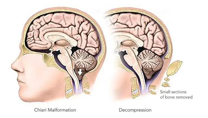
Chiari malformation surgery, also known as posterior fossa decompression surgery, is performed to relieve pressure on the brain and spinal cord caused by displaced brain tissue. Surgery is generally recommended when symptoms become severe, neurological problems worsen, or imaging shows significant compression of the cerebellum or disruption of cerebrospinal fluid (CSF) flow. Most patients experience meaningful symptom improvement after surgery, particularly when treated by an experienced neurosurgeon.
Understanding Chiari Malformation
A Chiari malformation is a neurological condition in which part of the cerebellum extends through the opening at the base of the skull (foramen magnum). This abnormal positioning can place pressure on the brainstem and spinal cord while interfering with the normal circulation of cerebrospinal fluid.
Not every person diagnosed with Chiari malformation requires surgery. Many individuals have mild symptoms or discover the condition incidentally during MRI scans performed for unrelated reasons. Others may develop progressively worsening symptoms that significantly affect their daily lives, making surgical treatment the most appropriate option.
At Advanced Neurosurgery Associates, our board-certified neurosurgeons evaluate each patient carefully to determine whether conservative treatment or surgical intervention offers the best outcome.
What Are the Symptoms of Chiari Malformation?
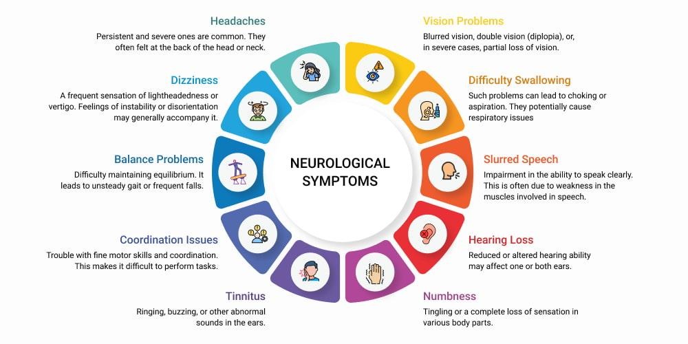
Symptoms vary depending on the severity of the condition and the degree of pressure placed on surrounding nerves and brain structures.
Common symptoms include:
- Severe headaches, especially after coughing or straining
- Neck pain
- Balance and coordination problems
- Dizziness or vertigo
- Numbness or tingling in the hands or feet
- Muscle weakness
- Difficulty swallowing
- Blurred or double vision
- Ringing in the ears (tinnitus)
- Sleep disturbances
- Difficulty with fine motor skills
Some patients experience symptoms gradually over many years, while others notice a sudden worsening after trauma or increased pressure inside the skull.
Learn more: Chiari Malformation Symptoms
Understanding the Different Types of Chiari Malformation
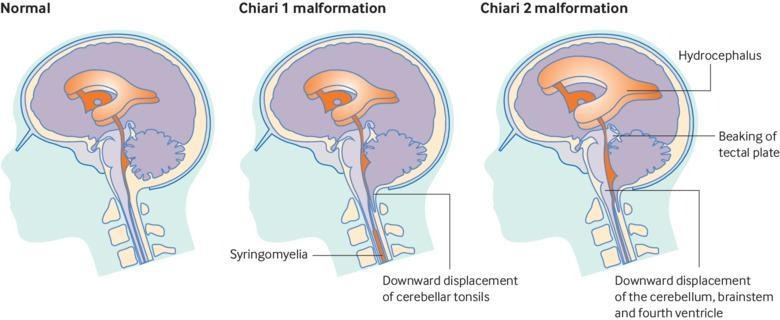
Knowing the type of Chiari malformation helps determine the most effective treatment approach.
Type I Chiari Malformation
This is the most common form and often develops during childhood or adulthood. Symptoms may not appear until later in life when pressure on the brain increases.
Many patients with Type I benefit from posterior fossa decompression surgery if neurological symptoms continue to worsen.
Type II Chiari Malformation
Usually diagnosed during infancy, Type II is commonly associated with spina bifida and hydrocephalus. Treatment often requires collaboration between pediatric neurosurgeons and other specialists.
Type III Chiari Malformation
A rare and severe form in which brain tissue protrudes through a defect in the skull. Early surgical treatment is usually necessary.
Type IV Chiari Malformation
This extremely rare type involves incomplete development of the cerebellum and requires individualized treatment planning.
Read our detailed guide: Chiari Malformation Types Explained: Type 1 vs Type 2 vs Type 3 vs Type 4
How Is Chiari Malformation Diagnosed?
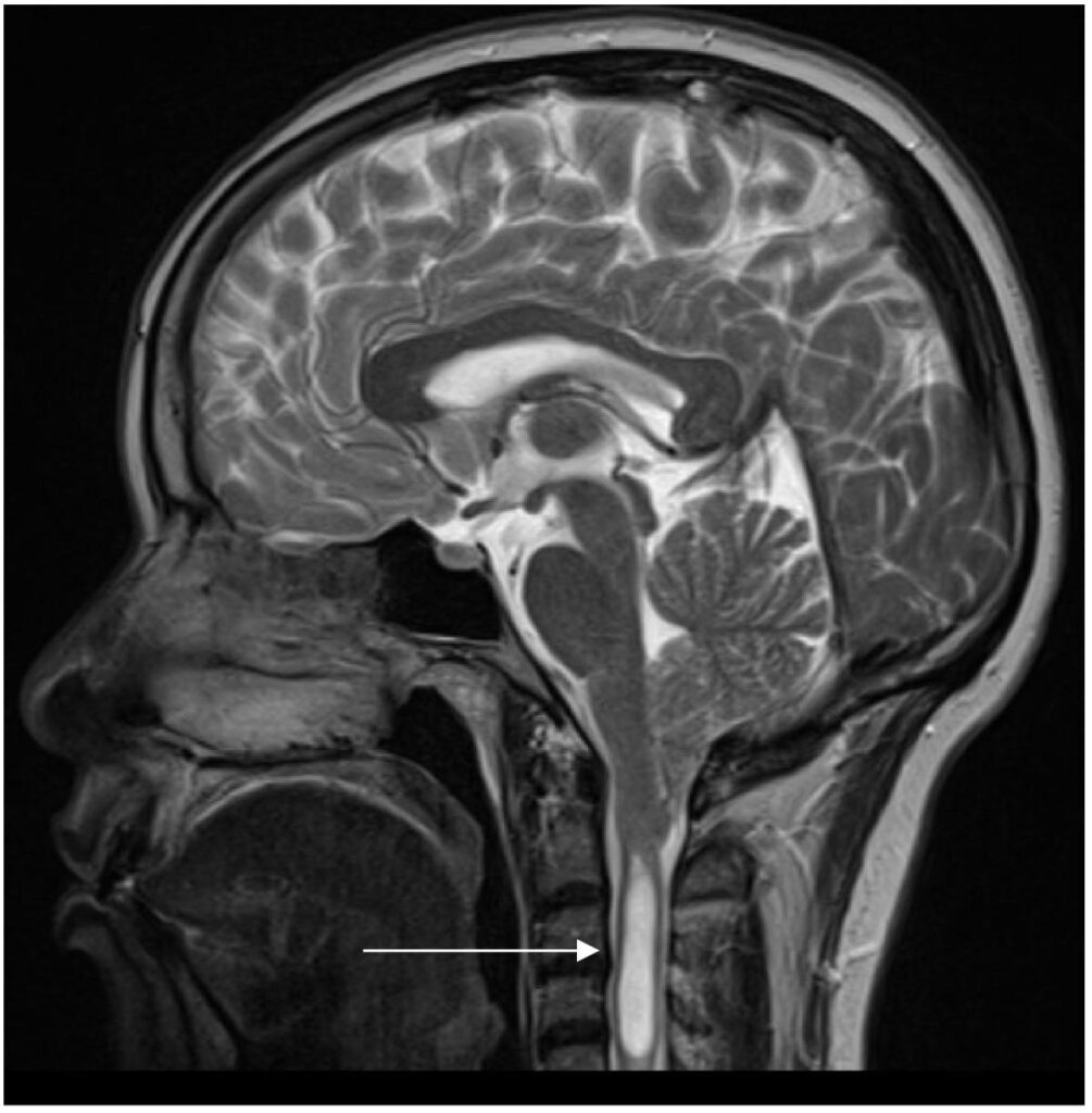
A thorough evaluation is essential before recommending surgery. Your neurosurgeon may perform:
Neurological Examination
This assessment evaluates muscle strength, reflexes, balance, sensation, coordination, and cranial nerve function.
MRI Scan
MRI is the gold standard for diagnosing Chiari malformation. It provides detailed images of the brain, cerebellum, spinal cord, and cerebrospinal fluid pathways.
MRI also helps identify associated conditions such as:
- Syringomyelia
- Hydrocephalus
- Tethered cord syndrome
- Brainstem compression
Cine MRI
Some patients benefit from Cine MRI, which evaluates the movement of cerebrospinal fluid around the brain and spinal cord. Reduced CSF flow often indicates that decompression surgery may be beneficial.
CT Scan
Although MRI is preferred, CT imaging may be used to assess skull anatomy before surgery.
When Is Chiari Malformation Surgery Needed?

Not every patient requires surgery. Many individuals with mild symptoms can be managed through regular monitoring and conservative treatment.
However, surgery may be recommended if you experience:
- Persistent or worsening headaches
- Progressive weakness or numbness
- Difficulty walking or maintaining balance
- Swallowing problems
- Sleep apnea related to brainstem compression
- Syringomyelia
- Significant blockage of cerebrospinal fluid flow
- Progressive neurological deficits
- Symptoms that interfere with daily activities despite conservative treatment
The primary goal of surgery is to relieve pressure on the brain, restore normal CSF circulation, and prevent permanent neurological damage.
Can Chiari Malformation Be Treated Without Surgery?
Yes. If symptoms are mild and imaging does not show significant compression, your neurosurgeon may recommend non-surgical management, including:
- Pain medications
- Physical therapy
- Activity modifications
- Regular neurological evaluations
- Periodic MRI monitoring
However, conservative treatment does not correct the underlying structural abnormality. If symptoms progress, surgery may become the most effective long-term solution.
Preparing for Chiari Malformation Surgery
Before recommending surgery, your neurosurgeon will review:
- MRI findings
- Neurological examination results
- Overall health
- Previous treatments
- Severity of symptoms
- Impact on your quality of life
Your healthcare team may also order:
- Blood tests
- Cardiac evaluation
- Anesthesia consultation
- Updated imaging studies
Preparing thoroughly helps reduce surgical risks and supports a smoother recovery.
How Is Chiari Malformation Surgery Performed?
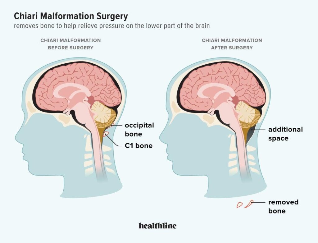
Once your neurosurgeon determines that surgery is the best treatment option, the most common procedure is posterior fossa decompression surgery. This operation is designed to create more space at the base of the skull, relieve pressure on the cerebellum and brainstem, and restore the normal flow of cerebrospinal fluid (CSF).
What Happens During the Procedure?
The exact surgical approach depends on your anatomy and the severity of the Chiari malformation, but most procedures follow these steps:
- General anesthesia is administered to ensure you are asleep and comfortable.
- A small incision is made at the back of the head.
- The neurosurgeon removes a small portion of bone from the lower part of the skull (posterior fossa) to create additional space.
- In some cases, part of the first cervical vertebra (C1) may also be removed.
- If needed, the protective covering of the brain (the dura) is opened and enlarged using a surgical patch (duraplasty) to improve CSF flow further.
- The incision is closed, and you are moved to recovery for monitoring.
The surgery typically takes 2–4 hours, depending on the complexity of the case.
Benefits of Chiari Malformation Surgery
The primary goal of surgery is not simply to reduce pain—it is to prevent further neurological damage and improve overall quality of life.
Many patients experience improvements in:
- Headaches
- Neck pain
- Balance and coordination
- Numbness and tingling
- Muscle weakness
- Difficulty swallowing
- Sleep disturbances
- Dizziness
Patients with syringomyelia may also experience stabilization or improvement after restoration of normal CSF flow.
What Are the Risks of Chiari Malformation Surgery?
Like any brain or spine procedure, Chiari malformation surgery carries potential risks. Fortunately, serious complications are uncommon when performed by an experienced neurosurgeon.
Possible risks include:
- Infection
- Bleeding
- Cerebrospinal fluid (CSF) leak
- Blood clots
- Wound healing problems
- Reaction to anesthesia
- Persistent headaches
- Temporary neurological symptoms
- Need for additional surgery in rare cases
Your neurosurgeon will discuss these risks and explain how they are minimized through careful planning and advanced surgical techniques.
Recovery After Chiari Malformation Surgery
Recovery varies from person to person, but most patients begin feeling better gradually over several weeks.
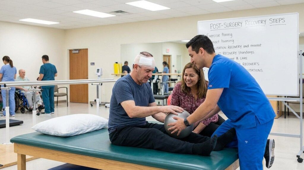
Week 1
- Hospital stay of 2–4 days
- Walking with assistance
- Pain managed with medication
- Rest and light activity
Weeks 2–4
- Increased walking
- Reduced discomfort
- Follow-up appointment
- Gradual return to normal daily activities
Weeks 4–8
- Many patients return to office work
- Improved balance and coordination
- Continued healing
After 3 Months
Most patients resume normal routines, although complete nerve healing may continue for several months.
Your neurosurgeon will provide personalized instructions regarding lifting, driving, exercise, and returning to work.
What Is the Success Rate of Chiari Malformation Surgery?
Posterior fossa decompression surgery has a high success rate when patients are appropriately selected.
Many published studies report that 70–90% of patients experience significant improvement in symptoms such as headaches, neck pain, balance problems, and numbness.
The outcome depends on several factors, including:
- Severity of symptoms before surgery
- Duration of nerve compression
- Presence of syringomyelia
- Overall health
- Surgical technique
- Postoperative rehabilitation
Early treatment often leads to better long-term outcomes.
Life After Chiari Malformation Surgery
Many patients return to work, school, sports, and other daily activities after recovery.
However, maintaining long-term neurological health is important.
Recommendations include:
- Attend scheduled follow-up visits.
- Complete all recommended MRI scans.
- Avoid activities that strain the neck until cleared by your surgeon.
- Follow physical therapy recommendations if prescribed.
- Report any new neurological symptoms promptly.
When Should You See a Neurosurgeon?
You should seek evaluation if you experience:
- Persistent headaches that worsen with coughing or straining
- Progressive numbness or weakness
- Difficulty walking or maintaining balance
- Difficulty swallowing
- Vision changes
- Chronic neck pain with neurological symptoms
- MRI findings suggesting Chiari malformation
Early diagnosis can help prevent permanent neurological complications and improve treatment outcomes.
Why Choose Advanced Neurosurgery Associates?
At Advanced Neurosurgery Associates, our board-certified neurosurgeons specialize in diagnosing and treating complex brain and spine conditions, including Chiari malformation.
We provide:
- Advanced diagnostic imaging
- Personalized treatment plans
- State-of-the-art surgical techniques
- Comprehensive postoperative care
- Compassionate, patient-centered treatment
Whether you require monitoring or surgery, our experienced team is committed to helping you achieve the best possible outcome
Frequently Asked Questions
Can Chiari malformation be cured with surgery?
Surgery cannot cure the structural abnormality but can effectively relieve pressure on the brain and restore CSF flow, significantly improving symptoms for many patients.
How long does Chiari malformation surgery take?
Most procedures take 2–4 hours, depending on the patient’s anatomy and surgical plan.
Is Chiari malformation surgery painful?
Some discomfort is expected after surgery, but pain is managed with medication and generally improves over the first few weeks.
How long is recovery?
Many patients return to light activities within 4–8 weeks, while complete healing may take several months.
Will I need physical therapy?
Some patients benefit from physical therapy to improve balance, strength, and mobility during recovery.
Can Chiari malformation return after surgery?
Most patients experience lasting improvement, although some may require additional evaluation if symptoms recur.
Is surgery always necessary?
No. Why Choose Advanced Neurosurgery Associates? are often managed with observation and periodic imaging.